Heart disease: 9 warning signs that appear on your skin
Warning signs of heart disease can appear on your skin and nails. If you notice any of the following signs, partner with a board-certified dermatologist or make an appointment to see your primary care doctor.
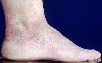
Swelling in your feet and lower legsWhat it may be telling you: Your heart isn’t working properly.
Many diseases of the heart cause fluid to build up in your feet and lower legs. As the fluid builds up, you may see swelling, which can extend as far as the upper legs and groin.
Medical name: Edema (medical term for swelling)
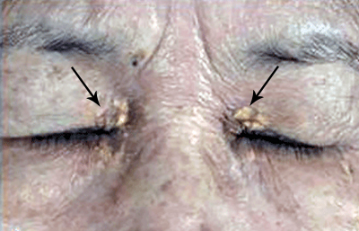
Yellowish-orange, waxy growths on your skin
What it may be telling you: You have unhealthy cholesterol levels and diabetes.
If you see yellowish-orange growths on your skin, you may have deposits of cholesterol under your skin. These painless deposits can appear in many areas, including the corners of your eyes, lines on your palms, or the backs of your lower legs.
If you notice these growths on any area of your skin, see your doctor. You may need cholesterol testing or another medical test. Unhealthy cholesterol levels require treatment, which can prevent life-threatening heart disease. Getting your cholesterol levels under control may also help clear the growths on your skin. If the growths don’t clear, a board-certified dermatologist can treat them.
Medical name: Xanthelasma (cholesterol deposits on the eyelids), Xanthoma (cholesterol deposit found elsewhere on the skin)
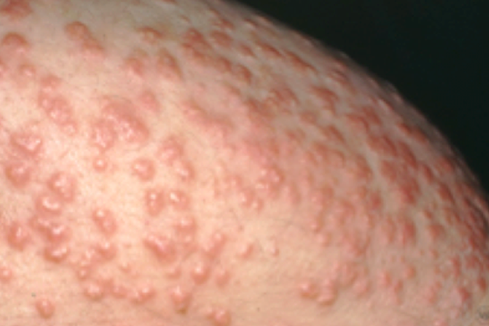
Clusters of waxy bumps that suddenly appear on your skin
What it may be telling you: You have skyrocketing cholesterol levels or diabetes.
The sudden appearance of these bumps can look like a rash, warts, or a contagious skin condition called molluscum contagiosum. These bumps are actually fatty deposits, which may be caused by extremely high levels of fats in the blood, especially triglycerides (a type of fat). A board-certified dermatologist knows the difference between these bumps and other skin conditions.
Treatment is essential to lower the triglycerides and treat any serious medical conditions, such as heart disease caused by the high cholesterol levels.
Medical name: Eruptive xanthoma (medical term for the sudden appearance of many fatty deposits)
Nails curve downward and the ends of your fingers are swollenWhat it may be telling you: For many people, these signs are harmless. They can also indicate a heart infection, heart disease, or lung problem.
If your fingers and nails look like this, it’s best to see your doctor to find out if you may have a medical condition, such as lung disease or a heart problem.
Medical name: Clubbing (term describes the downward turned nails and swollen fingers)
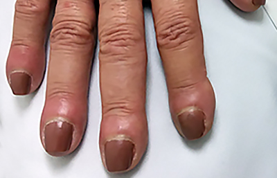
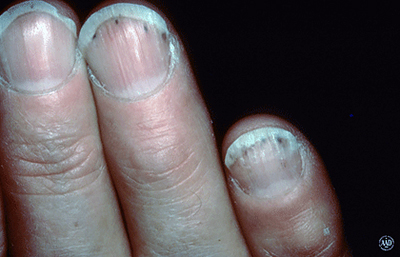
Brown, red or black lines under your nails What it may be telling you: Most people who see these lines under their nails have injured the nail in some way. If you cannot remember injuring your nail, see your doctor. These lines can be a sign of heart disease or another condition.
If you see brown, red, or black streaks that are short and involve the nails, sometimes it’s caused by an underlying heart disease, such as endocarditis (inflammation of the inner lining of the heart). If it’s a sign of heart disease, people tend to have symptoms, such as fever and a weak or irregular heartbeat.
Medical name: Splinter hemorrhage (line often looks like a splinter stuck under the nail)
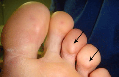
Painful lumps in your fingers, toes, or both What it may be telling you: You have an infection in your heart or blood vessels.
If you have a heart infection known as infective endocarditis, these painful lumps can develop in your fingers, toes, or both places. The lumps can last for a few hours to several days.
While the lumps go away on their own, you will need treatment for the infection. Bacteria cause this infection, so antibiotics can often treat it. Sometimes, surgery is also necessary.
Medical name: Osler nodes.
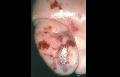
Brownish (or reddish) discoloration, usually on your sole(s) or palm(s) What it may be telling you: You have an infection in your heart or blood vessel.
These spots are painless and will clear without treatment, usually in a few days or weeks. However, the infection requires treatment.
Medical name: Janeway lesions
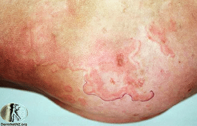
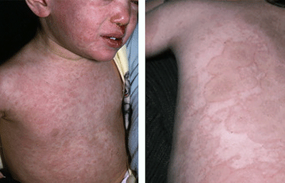
Non-itchy rash (flat spots with slightly raised edges) and fever What it may be telling you: You have rheumatic fever.
If your child develops strep throat, treating it quickly is important. When it’s not treated quickly, other medical problems can develop. One such problem is rheumatic fever. While this seldom happens in the United States today, rheumatic fever is common in developing countries.
A rash triggered by rheumatic fever can cause circular (or ring-like) marks that are bright pink or red and have raised borders and a pale center. This rash occurs on the arms, legs, and body, but usually not on the face. The lesions can last for a few hours and then reappear.
When a child has rheumatic fever, it can lead to lifelong heart disease. Rheumatic fever is a leading cause of heart disease in children.
Medical name: Erythema marginatum (name of the rash shown in this picture)
Rash and cracked, swollen lips that often bleed What it may be telling you: A child has Kawasaki disease.
When a child has a rash, fever, and extremely dry lips that may crack and bleed, Kawasaki disease may be the cause. This disease, which affects the blood vessels, usually develops in children between the ages of 6 months and 5 years of age.
While Kawasaki disease may go away on its own within 12 days without treatment, it damages the heart if left untreated.
Medical name: Mucocutaneous lymph node syndrome (another name for Kawasaki disease)
If you notice any of these signs, make an appointment to see your primary care doctor or a board-certified dermatologist. The sign could be harmless, but it’s important to get it checked out. Heart disease is easier to treat when found early.
Images
Images 1,3, 4, 5, 7: Used with permission of the American Academy of Dermatology National Library of Dermatologic Teaching Slides.
Images 2, 6, 9: Used with permission of Journal of the American Academy of Dermatology:
(2) J Am Acad Dermatol. 2017;77:728-34. (Fig 3A)
(6) J Am Acad Dermatol. 2009; 60(1):1-20.
(9) J Am Acad Dermatol. 2013;69:501.
Image 8: Used with permission of DermNet NZ. Last accessed May 11, 2018.
References
Hirschmann JV and Raugi GJ. “Blue (or purple) toe syndrome.” J Am Acad Dermatol. 2009; 60(1):1-20.
Khanna N, Roy A, et al. “Janeway lesions: an old sign revisited.” Circulation. 2013; 127(7):861.
Misin A, Di Bella S, et al. “Image of the month: ‘Diagnostic hands’: Janeway lesions.” Clin Med (Lond). 2017; 17(4):373-374.
Uliasz A and Lebwohl M. “Cutaneous manifestations of cardiovascular diseases.” Clin Dermatol. 2008; 26(3):243-54.