 $500.00
 $171.00


Conveniently pay your annual dues and take advantage of the many resources and benefits AAD membership offers.
Practical, dermatologist-specific products to help you in your day-to-day operations.
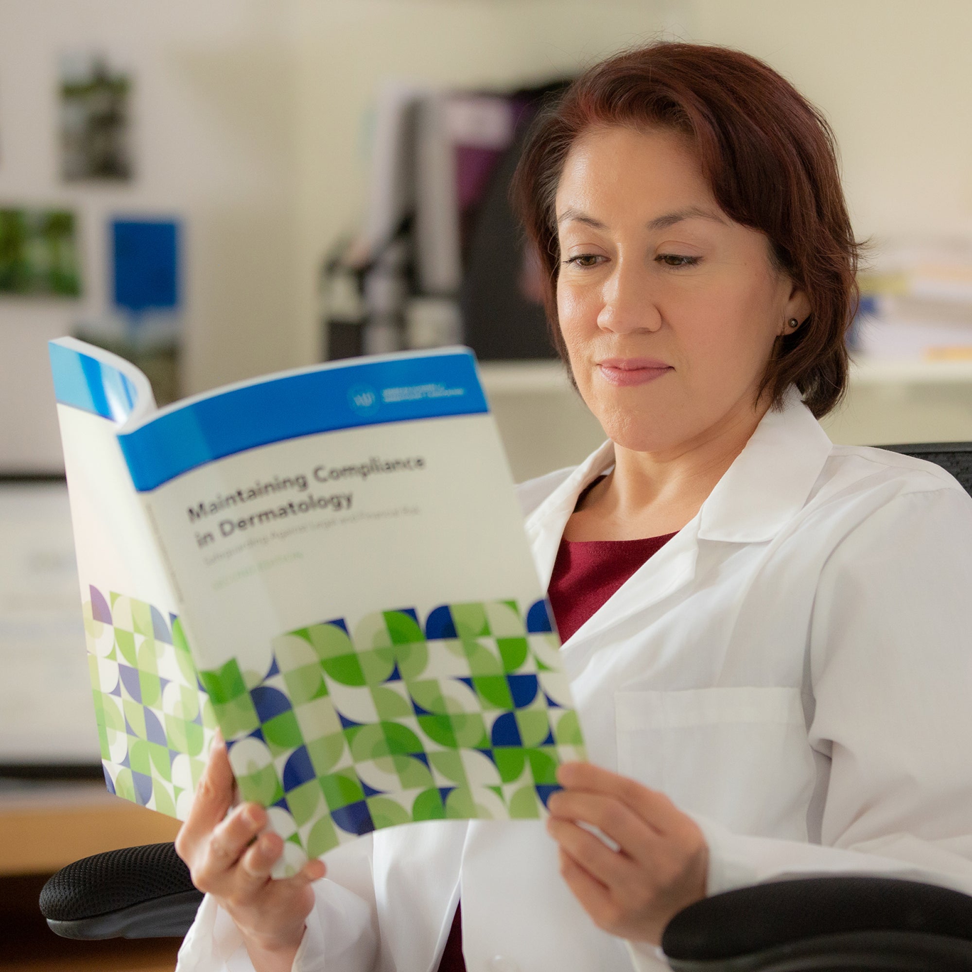
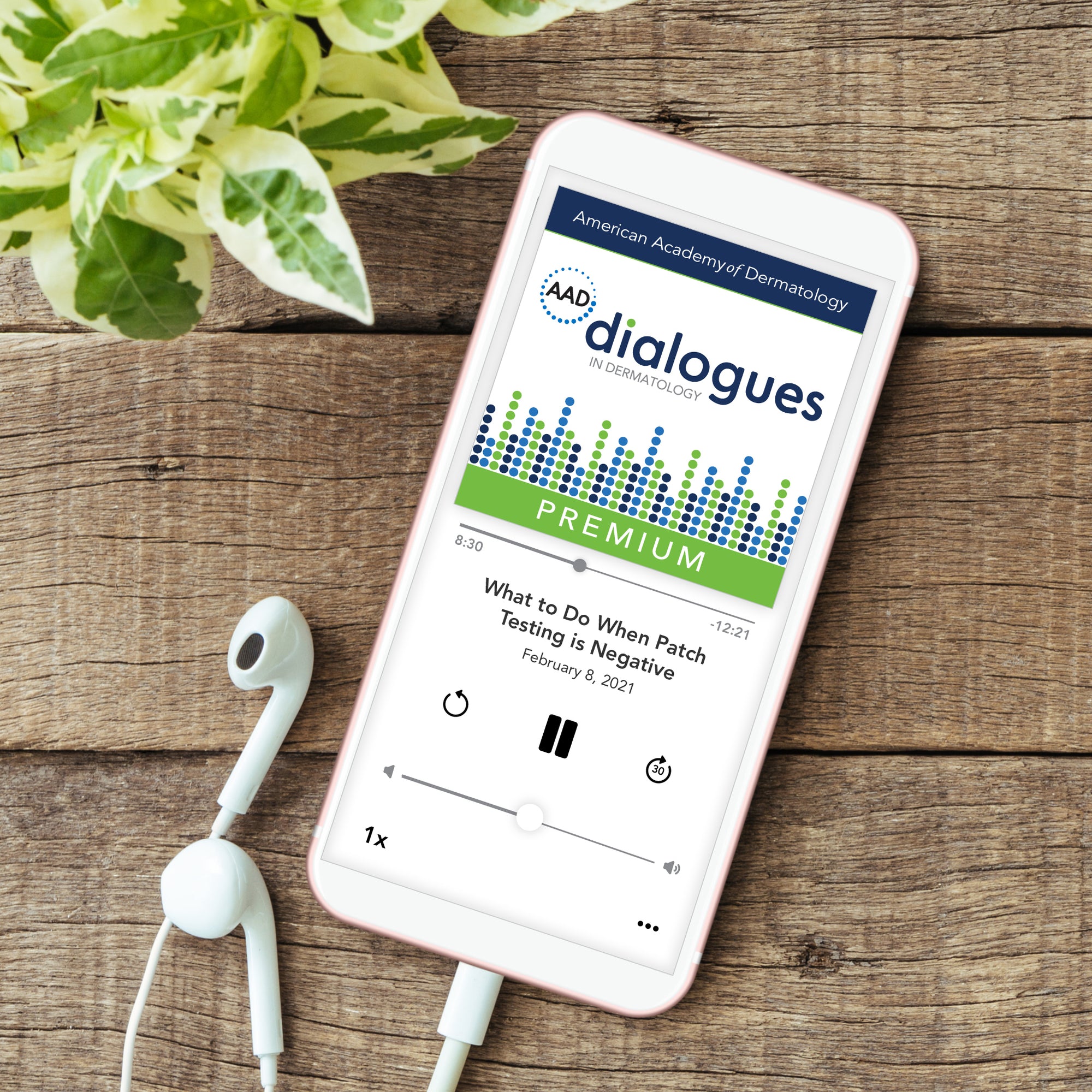
There’s no doubt about it. We have the best educational products to help you earn CME.
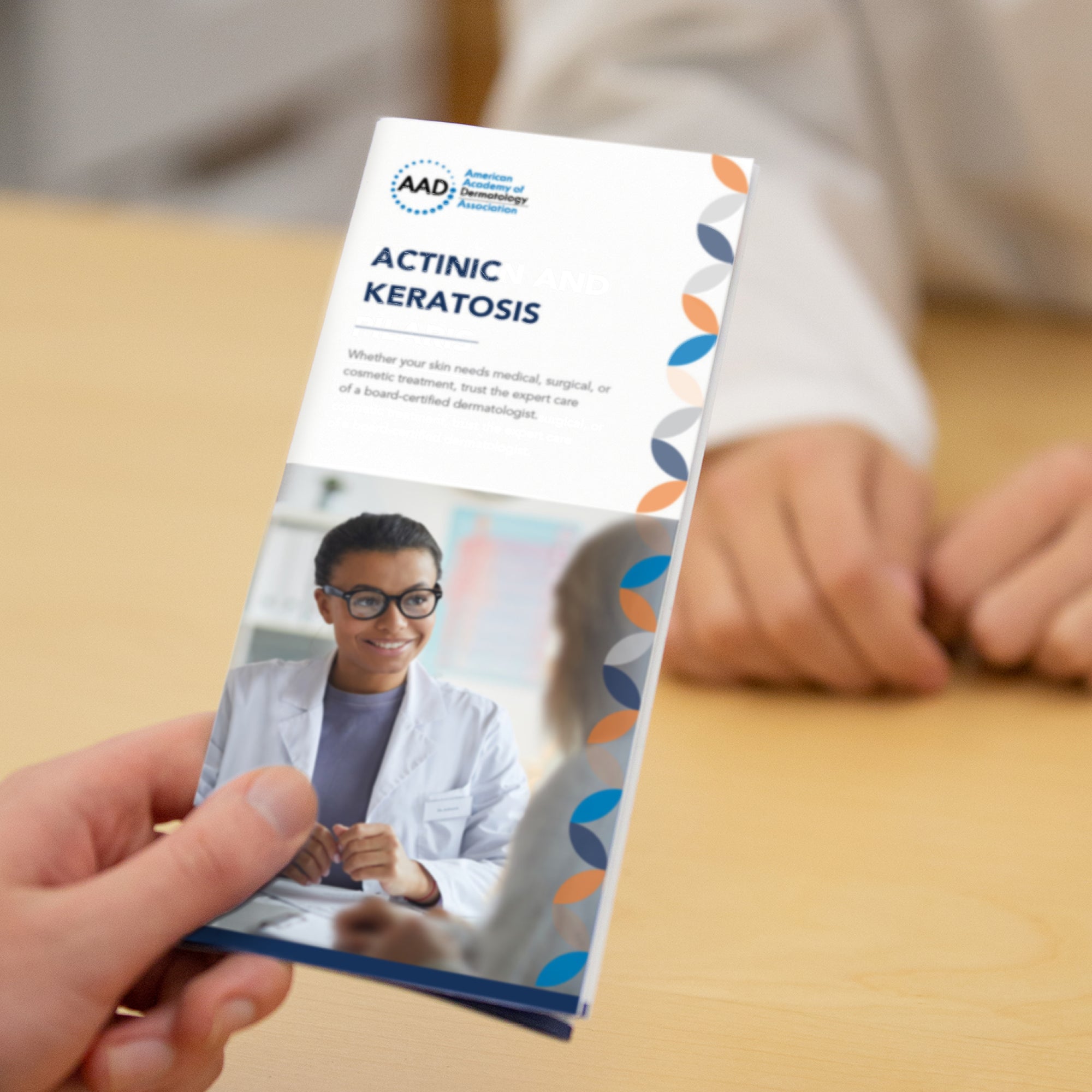
The best pamphlets on the market. Written by dermatologists for dermatologists. Why would you buy pamphlets anywhere else?
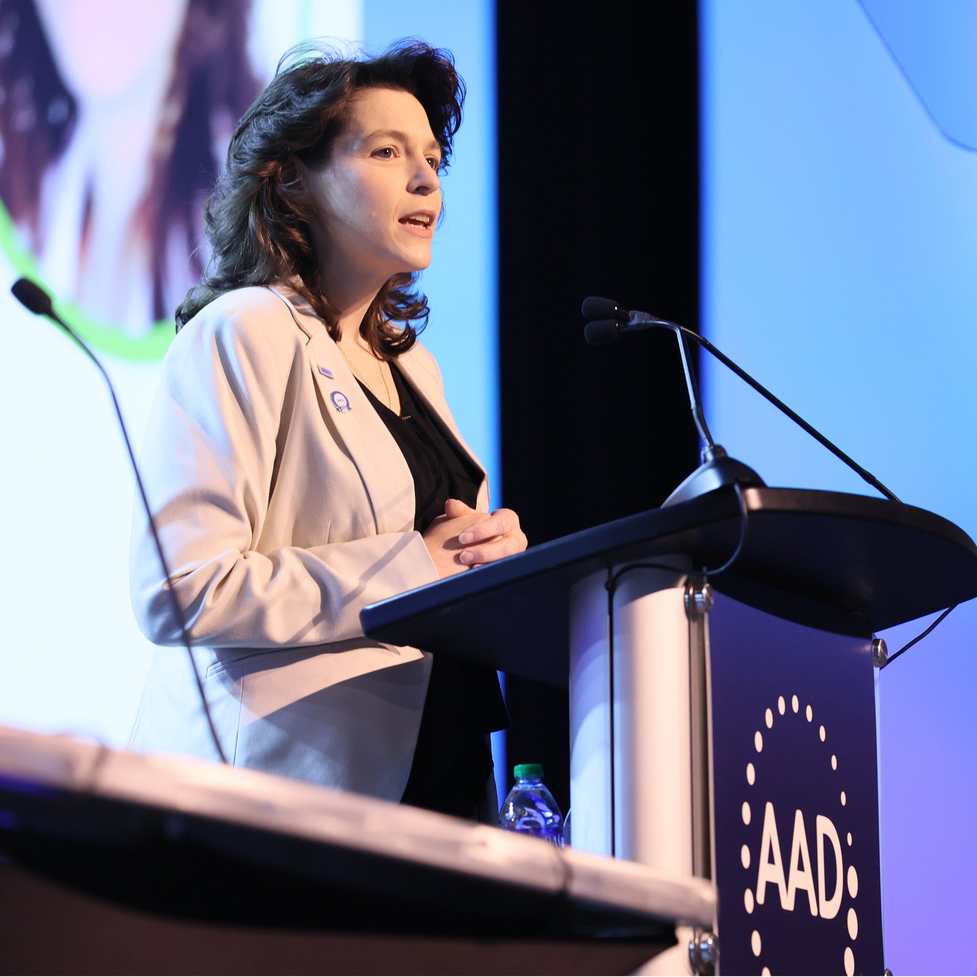
Discover our upcoming meetings and events.


Get in touch with our Member Resource Center with questions you may have on products, your membership, donations, whatever you can think of. We’re just waiting to hear from you.
Maybe you don’t want to talk to us. Don’t worry, we have these handy FAQs. We’ve got everything you need to know about returns, exchanges, shipping, and payments.